Scapular Dyskinesia Classification, Diagnosis, Management and Interventions
Overhead athletes have a higher prevalence of scapular dyskinesia of about 61% as compared with non-overhead athletes.
Symptoms of the isolated SICK scapula:
- Anterior shoulder pain (most common),
- Posterior superior scapular pain,
- Superior shoulder pain,
- Proximal lateral arm pain or any combination of above
Posterosuperior scapular pain may radiate into the ipsilateral paraspinous cervical region or the patient may complain of radicular/TOS type symptoms. The onset is almost always insidious.
Classification of Scapular Dysfunction
Type I or Inferior dysfunction. The primary external visual feature is the prominence of the inferior angle as a result of anterior tilting of the scapula in the sagittal plane. Inferior pattern presentation is better visualized while in the hands-on-hips position or during eccentric lowering from overhead elevation. According to Kibler, Type 1 pattern is most commonly found in patients with rotator cuff dysfunction.
Type 2 or Medial dysfunction. The primary external visual feature is the prominence of the entire medial scapular border due to internal rotation of the scapula in the transverse plane. As with Type 1, the Type 2 presentation becomes more evident in the hands-on-hips position and during active eccentric lowering from overhead. Medial pattern dysfunction most often occurs in patients with glenohumeral joint instability.
Type 3 or Superior dysfunction. Characterised by excessive and early elevation of the scapula during UE elevation. This pattern has been referred to as compensatory shoulder hiking or shrug and is most often seen in patients with rotator cuff dysfunction and deltoid-rotator cuff force couple imbalances.
Diagnostic Procedures
Current tests and measures, while proven to be reliable, have not altogether shown strong validity by demonstrating correlation with biomechanical motion, symptoms, pathology, or outcomes.
Symptom Altering Tests
- Scapular retraction test: Baseline AROM and pain is evaluated. This test is positive if pain is reduced as the therapist assists active elevation by applying a posterior tilt and external rotation motion to the scapula. This application may be used in conjunction with other tests such as Neer’s, Hawkin’s-Kennedy, and Jobe’s relocation.
- Scapular assistance test: Baseline AROM and pain is evaluated. The therapist then applies an assist to scapular dynamics. This test is positive if ROM is increased or pain is reduced as the therapist manually assists scapular upward rotation during active UE elevation.
- Lateral scapular Slide test (LSST)]: Measurements are taken from spine of scapulae to T2/T3, Inferior angle of scapulae to T7/T9 and superior angle of scapulae to T2. The measurements are taken in 3 positions, (A) sitting/standing with arms resting on the side, (B) Hands on the waist, Thumbs Posteriorly (45 abduction), (C) 90 degrees abduction and maximal internal rotation. Measurement should not vary more than 1 to 1.5 cm, more the 1.5 cm difference significant.
- Isometric Scapular Pinch test: Patient in standing position and is asked to actively squeeze or retract the scapulae together as hard as possible. Normal Response: An individual able to hold the squeeze or 15 to 20 sec without any burning pain or noticeable weakness. Positive: Burning pain present. Watch for: patient relaxing the contraction.
- Scapular Load test: As in position (2) for LSST, Manual load is applied in anterior, posterior, inferior or superior direction to the arm, and scapula should not move more than 1.5 cm.
- Wall pushup test: Patient performs wall pushups for 15 to 20 times. Weakness of scapular muscles (mainly serratus anterior) or winging usually shows up with 5 to 10 pushups. For stronger or younger population, perform the test on floor.
Manual Muscle Testing
MMT for the middle and lower trapezius, and serratus anterior.
Pectoralis Minor Muscle Tightness
Current measures examine mm length at resting positions, not at maximal length. Unfortunately, there are no validated clinical measures to identify a patient as having a tight pectoralis minor muscle.
Static Measurements
Static measurements consist of (1) infera: difference in vertical height of the superomedial scapular angle of the dropped scapula compared to contralateral angle. (2) Lateral displacement: difference of superomedial scapular angle from midline (3) abduction: difference in angular degrees of medial scapular margin from plumb line.
Management / Interventions
Intervention is aimed at and pectoralis minor restriction and restoring periscapular mm balance through exercises promoting early and increased serratus anterior, lower, and middle trapezius activation while minimising upper trapezius activity.
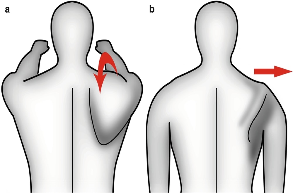
- Manual grade 4 mobilization to reduce posterior capsule tension, cross-body stretch.
- Manual stretching and soft tissue mobilization to decrease pec minor tension (cadaveric studies imply that a position of 150 degrees elevation with 30 degrees scapular retraction is optimal).
- Exercises of sidelying forward flexion, external rotation, prone extension, and prone horizontal abduction to strengthen middle and lower trapezius over upper trapezius.
- Quadruped and variable push-up positions to activate serratus anterior.
Rehab Protocol
Phase I
Flexibility
- Soft tissue Release: Pec Release, Posterior RTC release, Posterior capsule Release, Upper traps and levator scapulae release (Manual/self-release techniques)
- Tspine Mobility (manual Mobs/Manips)
- Tspine Extension Ex
- Pec Stretch
- Sleeper’s stretch
- Genie Stretch
- Upper traps stretch
Isometrics
- Scapular Pinches
- Robbery Pinches
- Low Row Wall isometrics
- Shoulder ER isometrics
- Scapular Depressor isometrics
Isotonics
- Scapular Pinches w/ Theraband
- Low Row w/ Theraband
- Shoulder ER/IR in standing
- Dynamic Hug
- Scapular punches
- Cheerleader Exercises.
Phase II
- Seated Rows
- High Rows
- Prone Rows
- Standing Robbery w/ theraband
- Prone Y, I, T and W
- Lawnmower/Standing D2 Cocking
- Side lying ER
- PNF D1/D2
- Lats Pull downs
- Scapular clocks
- Wall washes
- Thrower’s 10
- Manually Resisted scapular strengthening
- Stretches
Phase III
CKC exercises and advanced exercises.
- Super 6: Upright Row, Dynamic Hug, Cocking/Decelaration, Cocking/acceleration, B/L D2, B/L pullovers
- Standing cable column punches
- Bear crawl on Swiss ball
- Plyoball Decelaration
- Seated Pike lift
- Push Plus/Scapular Pushups
- Standing Snow Angels
- Wall ball Scours
- Stretches
References
This article was written by the Physiopedia site. You can read the original text here.