Things You May Need To Know About Ankle Sprain
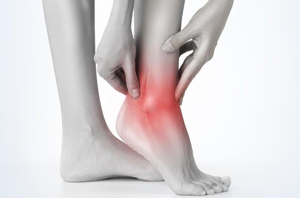
An ankle sprain is a common injury. Inversion-type, lateral ligament injuries represent approximately 85% of all ankle sprains. The incidence of ankle sprain is highest in sports populations. Poor rehabilitation after an initial sprain increases the chances of this injury recurrence. The ankle joint is the body part that is second most likely to be injured in sport. In the United States of America, the total cost of ankle sprains is approximately $2 billion. A meta-analysis by Doherty et al, found that indoor sports carry the greatest risk of ankle sprain with an incidence of 7 per 1,000 cumulative exposures.
Risk Factors
Several intrinsic and extrinsic risk factors predispose an athlete to chronic ankle instability. The most common risk factor is the previous history of the sprain. A previous sprain may compromise the strength and integrity of the stabilizers and interrupt sensory nerve fibers. Sex, height, weight, limb dominance, postural sway, and foot anatomy are intrinsic. Extrinsic risk factors may include taping, bracing, shoe type, competition duration and intensity of activity.
Differential Diagnosis
- The patient presents with inversion injury or forceful eversion injury to the ankle. May have a previous history of ankle injuries or instability.
- Able to partial weight-bear only on the affected side.
- If a patient presents with a description of the cold foot or paraesthesia, suspect neurovascular compromise of the peroneal nerve.
- Tenderness, swelling, and bruising can occur on either side of the ankle.
- No bony tenderness, deformity, or crepitus present.
- Passive inversion or plantar flexion with inversion should replicate symptoms for a lateral ligament sprain. Passive eversion should replicate symptoms for a medial ligament sprain.
- Special Tests: +ve Anterior Draw, Talar Tilt or Squeeze Test (depending on the structures involved)
Differential Diagnosis
- Impingement
- Tarsal Tunnel Syndrome
- Sinus Tarsi Syndrome
- Cartilage or osteochondral injuries
- Peroneal Tendinopathy or subluxation
- Posterior Tibial Tendon Dysfunction
Clinical Examination
With an ankle sprain, multiple structures may be involved, therefore a full foot and ankle assessment is recommended, including the mechanism of injury, observation of the patient’s gait pattern, standing posture and wear on the individual’s shoes. Any gross deformity, mal-alignment or atrophy of the musculature should also be observed and noted as well as any edema and/or ecchymosis.
There are numerous grading systems used for the classification of ligament sprains, each having its strengths and weaknesses. Different therapists may employ different systems so effective continuity of care, the patient should see the same therapist each time. Authors do not always disclose which system they used, reducing the rigor and quality of some research.
The traditional grading system for ligament injuries focuses on a single ligament
- Grade I represents a microscopic injury without stretching of the ligament on a macroscopic level.
- Grade II has macroscopic stretching, but the ligament remains intact.
- Grade III is a complete rupture of the ligament.
As there are ankle multiple ligaments across the joint it may not be always straightforward to use a grading system that is designed for describing the state of a single ligament unless it is certain that only a single ligament is injured. Some authors have therefore resorted to grading lateral ankle ligament sprains by the number of ligaments injured. It is, however, hard to be certain of the number of ligaments torn unless it is clear, high-quality radiographic or surgical evidence.
A third system that can be adopted is a 3 graded classification based on the severity of sprain injury.
- Grade I Mild – Little swelling and tenderness with little impact on function
- Grade II Moderate – Moderate swelling, pain and impact on function. Reduced proprioception, ROM, and instability
- Grade III Severe – Complete rupture, large swelling, high tenderness loss of function and marked instability
This scale is largely subjective due to individual therapist interpretation. However, the same can be said for the other classifications unless clear radiographic evidence is available or assessed and treated by surgical intervention.
Palpation is used to feel for the structures that may be involved in the injury, including bone, muscle, and ligamentous structures, followed by an active and passive range of movement assessment.
- Anterior Draw – tests the ATFLSpecial Tests
- Talar Tilt – tests the CFL
- Posterior Draw – tests the PTFL
- Squeeze test – for syndesmotic sprain
- External rotation stress test (Kleiger’s test) – syndesmotic sprain
It is recommended that these tests be performed at 4-7 days post-acute injury to allow the initial swelling and pain to settle, enabling the therapist to gain a more accurate diagnosis.
Physical Therapy Management
Mild Ankle Sprain
- Natural full recovery within 14 days
- Taping and follow up to evaluate healing progression
First-time lateral ligament sprains can be innocuous injuries that resolve quickly with minimal intervention and some approaches suggest that only minimal intervention is necessary. The NICE guidelines 2016 recommend advice and analgesia, but not routine physiotherapy referrals. However, it has also been highlighted that the recurrence rate of first-time lateral ankle sprains is 70%. With the recurrence rate so high and the guidelines not recommending any rehabilitation, this approach has been questioned.
Severe Ankle Sprain
Physiotherapy is required with functional therapy of the ankle shown to be more efficient than immobilization. Functional therapy treatment can be divided in 4 stages, moving onto the next stage as tissue healing allows
- Inflammatory phase,
- Proliferative phase,
- Early Remodelling,
- Late Maturation and Remodelling.
Inflammatory Phase (0-3 days)
Reduction of pain and swelling and improve circulation and partial foot support
The most common approach to manage ankle sprain is the PRICE protocol: Protection, Rest, Ice, Compression, and Elevation
Recommendations for the Patient:
- Protection: Protect the ankle from further injury by resting and avoiding activities that may cause further injury and/or pain
- Rest: Advise rest for the first 24 hours after injury, possibly with crutches to offload the injured ankle and altering work and sport and exercise requirements as needed
- Ice: Apply a cold application (15 to 20 minutes, one to three times per day)
- Compression: Apply a compression bandage to control swelling caused by the ankle sprain
- Elevation: Ideally elevate the ankle above the level of the heart, but as a minimum, avoid positions where the ankle is in a dependent position relative to the body
Despite its widespread clinical use, the precise physiologic responses to ice application have not been fully elucidated. Moreover, the rationales for its use at different stages of recovery are quite distinct. There is insufficient evidence available from RCTs to determine the relative effectiveness of RICE therapy for acute ankle sprains in adults. But no evidence exists to reject the RICE protocol.
Foot and Ankle ROM
- The patient performs active movements with the toes and ankle within pain-free limits to improve local circulation.
- Manual therapy in the acute phase could also effectively increase ankle dorsiflexion.
- Anteroposterior manipulation and RICE results in greater improvement in range of movement than the application of RICE alone.
Proliferative Phase (4-10 days)
Recovery of foot and ankle function and improved load-carrying capacity.
1. Patient education regarding the gradual increase in activity level, guided by symptoms.
2. Practise Foot and Ankle Functions
- Range of Motion
- Active Stability
- Motor Coordination
It is important to begin early with the rehabilitation of the ankle. First-week exercises produce significant improvements to short-term ankle function.
3. Tape/Brace :
- Apply tape as soon as the swelling has decreased.
- Tape or a brace use depends on patient preference
Early Remodelling (11 -21 days)
Improve muscle strength, active (functional) stability, foot/ankle motion, mobility (walking, walking stairs, running).
Practice foot and ankle functions
- Provide information about possible preventive measures (tape or brace)
- Advice regarding appropriate shoes to wear during sports activities, in relation to the type of sport and surface
- Practice balance, muscle strength, ankle/foot motion and mobility (walking, stairs, running).
- Look for a symmetric walk pattern.
- Work on dynamic stability as soon as loa-bearing capacity allows, focusing on balance and coordination exercises. Gradually progress the loading, from static to dynamic exercises, from partially loaded to fully loaded exercises, and from simple to functional multi-tasking exercises. Alternate cycled with non-cycled exercises (abrupt, irregular exercises). Use different types of surfaces to increase the level of difficulty.
- Encourage the patient to continue practicing the functional activities at home with precise instructions regarding the expectations for each exercise.
- Advise wearing tape or a brace during physical activities until the patient is able to confidently perform static and dynamic balance and motor coordination exercises.
Late Remodelling and Maturation
Improve the regional load-carrying capacity, walking skills and improve the skills needed during activities of daily living as well as work and sports.
- Practice motor coordination skills while performing mobility exercises
- Continue to progress the load-bearing capacity as described above until the pre-injury load-carrying capacity is reached
- Increase the complexity of motor coordination exercises in varied situations until the pre-injury level is reached
- Encourage the patient to continue practicing at home
Chronic Ankle Instability
On-going issues following a lateral ligament injury within the ankle are reported in 19-72% of patients. An ability to complete certain movement tasks, evidence of deficits during the Star Excursion Balance Test and self-reported function as quantified using the Foot and Ankle Ability Measure can be utilized as predictive measures of a Chronic Ankle Instability (CAI) outcome in the clinical setting for patients with a first time lateral ankle sprain injury. Around 20% of people develop CAI and this has been attributed to a delayed muscle reflex of stabilizing lower leg muscles, deficits in lower leg muscle strength, deficits in kinaesthesia or impaired postural control.
Chronic ankle instability has been describing as a combination of mechanical (pathological laxity, arthrokinematic restrictions, and degenerative and synovial changes) and functional (Impaired proprioception and neuromuscular control, and strength deficits) insufficiencies. A sound treatment program must adhere to both mechanical and functional inefficiencies.
It is recommended that all patients undergo conservative treatment to improve stability and improve the muscle reflex and strength of the lower limb stabilizing muscles. Although this will help some individuals, it cannot compensate for the deficit of the lateral ligament complex, and surgery is occasionally required.